

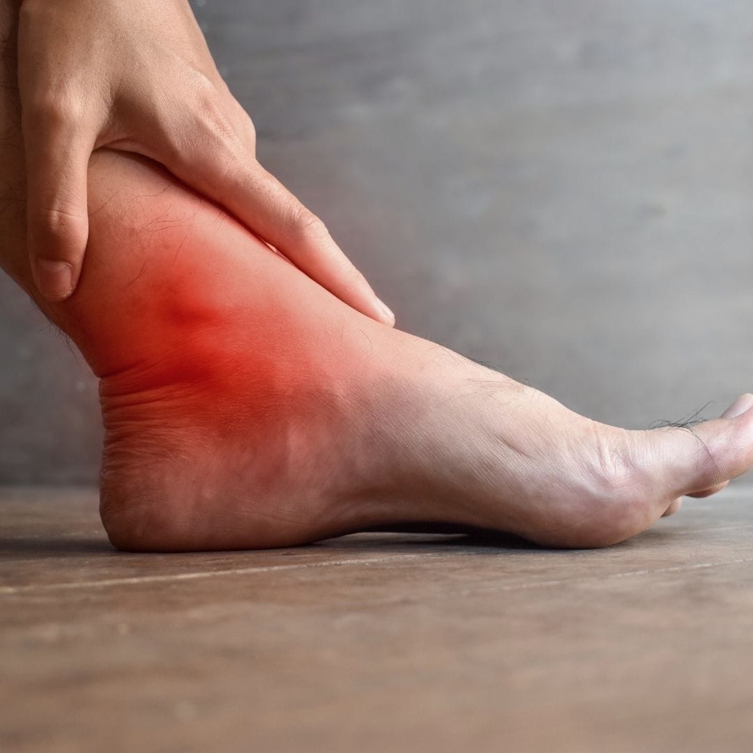
Almost everyone has rolled an ankle at some point. The immediate instinct is to rest for a few days, ice it, and assume it will sort itself out. For many Grade 1 sprains, this works. But for the larger, more significant sprains that go undertreated, the ligament heals incompletely — and the patient is left with an ankle that re-sprains easily, feels unstable on uneven ground, and never quite returns to how it felt before.
Ankle sprains are the most common sports injury in India — and the most undertreated. The same patient who would immediately seek a doctor for a broken bone will walk on a significant ankle ligament tear for weeks, assuming it just needs time. Understanding the grades, the proper treatment, and why incomplete healing matters is what separates a full recovery from years of chronic instability.
The Three Grades of Ankle Sprain
Mild ligament stretch, no tearing. Minimal swelling, point tenderness. Able to bear weight. Usually heals well with basic care.
Partial ligament tear. Moderate swelling and bruising. Some ankle instability. Needs structured physiotherapy for full recovery.
Complete ligament rupture. Significant swelling, bruising, instability. Cannot bear weight properly. May need surgical repair in active patients.
Most patients assume their ankle sprain is Grade 1. In reality, without clinical examination, there is no reliable way to grade a sprain — the amount of initial pain does not reliably predict severity. Some complete ligament ruptures are remarkably pain-free immediately after injury, only becoming symptomatic hours later when swelling develops.
Sprain or Fracture? — The Ottawa Ankle Rules
Ankle injuries frequently involve both ligament damage and bone injury — and distinguishing between them at the time of injury is not always possible without imaging. The Ottawa Ankle Rules are an evidence-based clinical tool that determines when X-ray is needed, developed specifically to avoid missing fractures that look like sprains.
Ottawa Ankle Rules — When to X-Ray
Ankle X-ray needed if
- Bone tenderness at back edge of the lower fibula (outer ankle)
- Bone tenderness at back edge of the lower tibia (inner ankle)
- Inability to bear weight immediately after injury AND in the emergency setting
Foot X-ray needed if
- Bone tenderness at the base of the 5th metatarsal (outer mid-foot)
- Bone tenderness over the navicular bone (inner mid-foot)
- Inability to bear weight immediately after injury AND in the emergency setting
The fifth metatarsal base fracture deserves special mention — it is one of the most commonly missed injuries in ankle trauma. The peroneal tendon attaches here, and the pull of this tendon during the sprain mechanism can avulse a fragment of bone. This injury looks and feels like a standard ankle sprain, but requires very different management. It is identified on foot X-ray, not ankle X-ray — which is why foot X-rays should be part of the assessment when mid-foot tenderness is present.
Why Ankle Sprains Become Chronic
Chronic ankle instability — a condition where the ankle continues to give way repeatedly, months or years after the original sprain — affects a significant proportion of people who have had moderate to severe ankle sprains. The underlying mechanism is almost always the same: inadequate rehabilitation of the original injury.
Incomplete Ligament Healing
Ligaments heal with scar tissue, not identical tissue. Without proper loading during healing, the scar tissue is mechanically inferior and the ankle remains structurally weak.
Lost Proprioception
Proprioception — the ankle's ability to sense its own position in space — is severely disrupted by ligament injury. Without specific balance retraining, this deficit persists even after pain resolves.
Muscle Weakness
The peroneal muscles on the outer leg are the primary dynamic stabilisers of the ankle. After a sprain, these muscles weaken from disuse and protective inhibition, leaving the joint mechanically unsupported.
Too Early Return to Sport
Returning to running, court sport, or heavy work before adequate ligament healing and proprioceptive retraining dramatically increases re-sprain risk — and each subsequent sprain causes additional ligament damage.

Symptoms That Need Assessment
Significant Swelling
Rapid swelling developing within minutes — suggests significant ligament or bone injury
Inability to Bear Weight
Cannot walk more than 4 steps without severe pain — warrants X-ray to rule out fracture
Ankle Giving Way
Ankle buckling repeatedly during walking — indicates instability from significant ligament laxity
Persistent Pain at 2 Weeks
Pain not improving after 2 weeks of basic rest and ice — warrants clinical review and possible imaging
Mid-Foot Tenderness
Pain over the outer mid-foot — possible fifth metatarsal fracture requiring specific X-ray
Previous Multiple Sprains
Repeated sprains of the same ankle — indicates chronic instability needing structured treatment
Treatment — POLICE Not RICE
The classic RICE protocol (Rest, Ice, Compression, Elevation) has been updated in modern sports medicine. The current evidence-based approach is POLICE: Protection, Optimal Loading, Ice, Compression, Elevation. The key change is "Optimal Loading" — complete rest is no longer recommended for ankle sprains. Early, controlled movement promotes better ligament healing than strict immobilisation.
Treatment pathway at Pure Ortho Hospitals, Sainikpuri
- Acute phase (Day 1-5): Protection from re-injury, ice and compression for swelling, elevation, early pain-free range of motion exercises to prevent stiffness
- Sub-acute phase (Week 2-4): Structured physiotherapy — peroneal muscle strengthening, progressive weight-bearing, balance and proprioception retraining
- Return to sport phase (Week 4-12): Sport-specific training, agility work, confirmation that balance is restored before full return
- Persistent instability: Lateral ligament reconstruction — surgical repair or reconstruction of the damaged ligaments using the Brostrom procedure for athletes with chronic ankle instability
- Associated injuries: Osteochondral lesions, peroneal tendon tears, or syndesmosis injuries identified on MRI are addressed as part of the treatment plan
Get Assessed If Your Ankle Sprain Involves
- Rapid significant swelling within 30 minutes of injury
- Inability to bear weight on the ankle after injury
- Bone tenderness at the inner or outer ankle bones or mid-foot
- Pain still significant after 2 weeks of rest
- Third or subsequent sprain of the same ankle
Call Pure Ortho Hospitals, Sainikpuri: 8686868208
Meet the Specialists at Pure Ortho Hospitals
Dr. G. Uday Sekhar Reddy
MBBS, MS Ortho, MCh Ortho
Dr. V.S. Abhilash Kumar S
MBBS, MS Ortho, FIJR, FISS (S.Korea, USA) — Clinical Director
Dr. Sai Karthikeya Badri
MBBS, D. Ortho, DNB
Dr. L. Sreeram
MPT (Ortho), FDOR, MIAP
Dr. L. Sri Dharani
BPT, MIAP, PTOTA (Canada)
Frequently Asked Questions
Other Departments at Pure Ortho Hospitals
A Properly Treated Ankle Sprain Heals Completely
An undertreated one becomes a lifetime of instability. Visit Pure Ortho Hospitals, Sainikpuri, Hyderabad — get the right grade assessment and the right rehabilitation plan.
Call 8686868208More from Pure Ortho Hospitals
This article is for patient education only. Please consult a qualified orthopaedic surgeon before making any treatment decisions.