


The patient in their 60s who used to walk a kilometre without stopping now has to sit down every 200 metres. They describe their legs becoming heavy, cramping, or numb — not from tiredness, but from a sensation that forces them to stop. Sitting down for a few minutes gives complete relief, and they can walk again — until the same thing happens again. Their back pain is manageable. It is the legs that are the problem.
This pattern is neurogenic claudication — the hallmark of lumbar spinal stenosis. It is one of the most recognisable presentations in spine medicine, and understanding why it happens — why walking produces symptoms and sitting relieves them — makes the diagnosis almost intuitive.
What Is Spinal Stenosis?
The spinal canal is the bony channel that runs through the centre of the vertebral column, housing the spinal cord and the nerve roots that branch off it. In a healthy spine this canal has adequate space for the neural structures inside. In spinal stenosis, this channel narrows — compressing the spinal cord, the cauda equina (the bundle of nerve roots in the lower spine), or the individual nerve roots as they exit through the lateral recesses and foramina.
Normal Spinal Canal
Adequate space around the nerve roots. Nerves pass freely through the canal and exit through the foramina without compression. No symptoms during normal activity.
Stenotic Spinal Canal
Canal narrowed by disc bulges, thickened ligament, or bone spurs. Nerve roots compressed — particularly during spinal extension (standing, walking). Symptoms with activity, relief with flexion (sitting).
Why Walking Causes Pain and Sitting Gives Relief
This is the key to understanding lumbar spinal stenosis — and it explains why the diagnosis is often missed for years when patients assume their leg symptoms are a circulation or muscle problem.
When you stand or walk, the lumbar spine extends (arches slightly backward). This extension naturally reduces the diameter of the spinal canal — the ligamentum flavum folds inward, and the posterior elements of the vertebrae move closer together. In a normal canal this is not an issue. In a stenotic canal, this extension pushes the already narrowed space to its limit, compressing the nerve roots and causing the characteristic leg symptoms.
When you sit, lean forward, or squat, the lumbar spine flexes. Flexion opens the canal — the ligamentum flavum tightens and pulls away from the canal, and the posterior elements separate slightly. Nerve root pressure is relieved. Symptoms disappear. This is why patients with spinal stenosis can often walk further when pushing a shopping trolley (the leaning posture keeps the spine flexed), and why cycling is typically painless while walking is not.
The Neurogenic Claudication Pattern
Neurogenic Claudication vs Vascular Claudication
Neurogenic (Spinal Stenosis)
Leg pain, cramping or numbness after walking. Relieved by sitting OR leaning forward. Can also cycle pain-free. Pulses normal. Symptoms bilateral. Worse going downhill (extension).
Vascular (Artery Disease)
Calf pain after walking. Relieved only by standing still — not by leaning. Cannot cycle. Reduced foot pulses. Usually one leg. Worse on any exertion regardless of posture.
Distinguishing between these two is critical — the treatments are completely different. If leg pain with walking goes away when you simply lean on a surface, or if you can cycle without pain but cannot walk the same distance, neurogenic claudication from spinal stenosis is the likely cause and spine evaluation is the right next step.
What Causes the Canal to Narrow?
In most patients over 50, spinal stenosis is the result of multiple overlapping degenerative changes rather than any single cause:
- Disc degeneration and bulging — degenerating discs lose height and bulge backward into the canal, reducing space from the front
- Ligamentum flavum hypertrophy — the ligament running along the back of the canal thickens with age, buckling inward and compressing the canal from behind — often the dominant cause of symptoms
- Facet joint arthritis and bone spurs — arthritic enlargement of the facet joints narrows the lateral recesses where nerve roots exit
- Spondylolisthesis — forward slippage of one vertebra on another further reduces canal diameter at that level
- Congenital narrow canal — some individuals are born with a smaller than average canal diameter and develop symptomatic stenosis earlier in life from smaller amounts of additional degeneration
Symptoms — The Full Picture
Neurogenic Claudication
Leg pain, heaviness, cramping or numbness after walking — relieved by sitting or leaning forward. The defining symptom of lumbar stenosis.
Reduced Walking Distance
The distance walked before symptoms onset gradually shortens over months and years as stenosis progresses.
Bilateral Leg Symptoms
Unlike a single disc prolapse causing one-sided sciatica, stenosis often affects both legs — though one side may be more affected than the other.
Back Pain
Present but often less prominent than leg symptoms. Back pain alone is not typical of stenosis — it is the leg symptoms with walking that are characteristic.
Difficulty Standing
Prolonged standing in one place — queuing, washing dishes — produces symptoms because the spine remains extended without the movement that walking provides some relief from.
Bladder Urgency
In severe central stenosis, cauda equina compression can cause urinary urgency or frequency — a more serious symptom requiring prompt evaluation.
Grades of Severity
Occasional Symptoms
Symptoms only with prolonged walking. Can walk 500m or more. No neurological deficit. Conservative management appropriate.
Frequent Limitation
Walking limited to 100-500m. Daily activities affected. Physiotherapy and injections often help. Surgery considered if not improving.
Severe Limitation
Walking less than 100m. Significant neurological deficit. Bladder symptoms may be present. Surgery usually recommended.
Diagnosis
Assessment at Pure Ortho Hospitals, Sainikpuri
- Clinical history — walking distance, posture dependence of symptoms, bladder symptoms, symptom duration and progression
- Neurological examination — reflexes, sensation, strength in both legs
- X-ray — shows disc height loss, spondylolisthesis, facet arthritis; does not show soft tissue compression directly
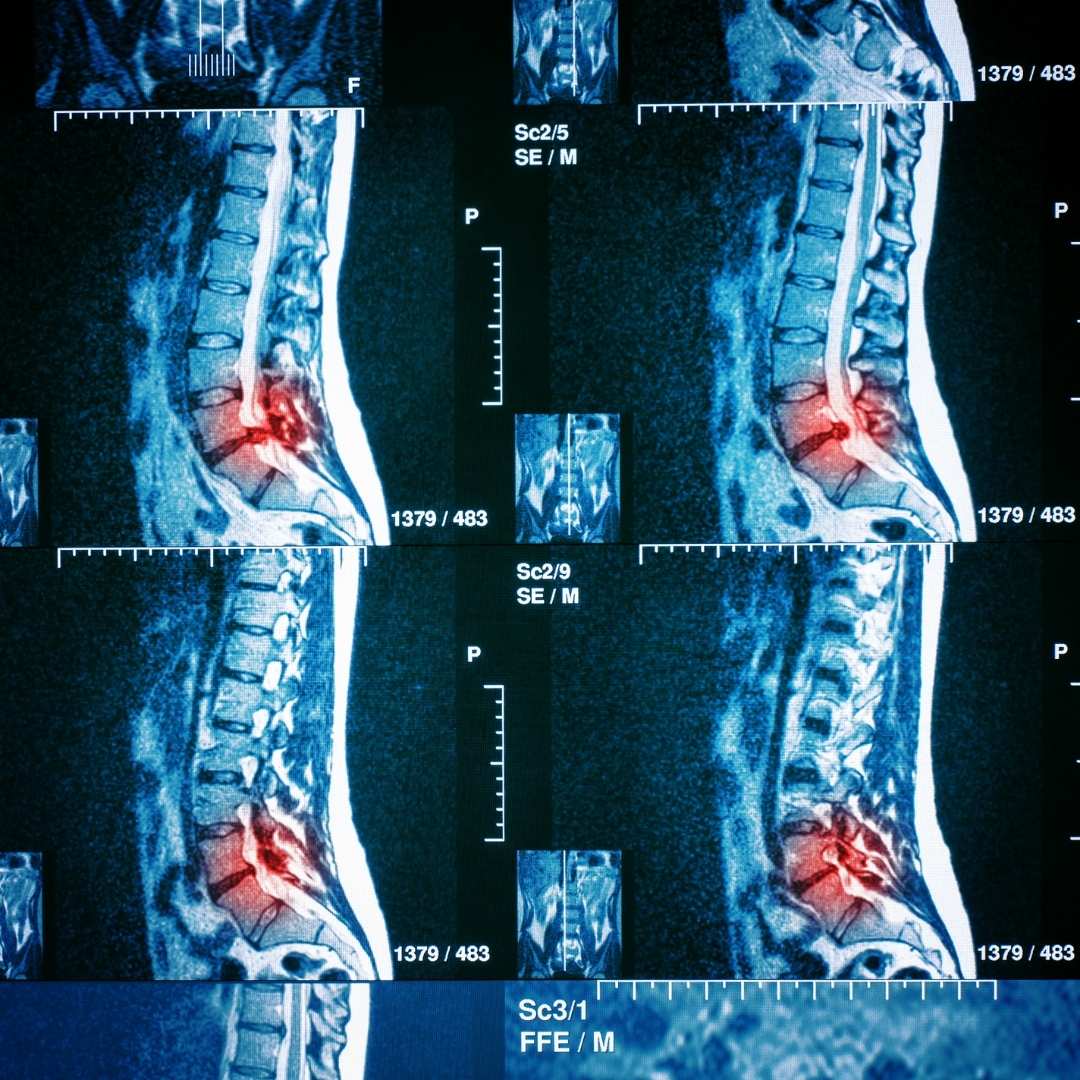
- MRI — the definitive investigation; shows the degree of canal narrowing, which structures are compressing the nerves, and how many levels are involved. Essential before any treatment decision.
- Vascular assessment — to exclude vascular claudication when clinical features overlap
- Dynamic films — flexion-extension X-rays to assess spinal instability when spondylolisthesis is present
Treatment — Conservative First
Conservative management options
- Physiotherapy — lumbar flexion exercises, core strengthening, postural training. Flexion-based exercise programmes maintain the spinal position that relieves canal pressure. Aquatic physiotherapy is particularly effective for patients who cannot walk far on land.
- Epidural steroid injections — corticosteroid injected into the epidural space around the compressed nerve roots reduces inflammation and swelling. Provides significant short to medium term relief in many patients. Can be repeated and helps confirm the level of pathology.
- Activity modification — cycling, swimming, and exercises in the flexed position maintain fitness without provoking symptoms. Walking with a slight forward lean (trolley walking technique) often allows greater distances.
- Pain management — neuropathic pain medications address the nerve-related burning and cramping component of symptoms.
Surgical options when conservative treatment is insufficient
- Laminectomy — removing the lamina (back arch of the vertebra) and thickened ligamentum flavum to directly widen the spinal canal. The standard and most effective procedure for central stenosis. Reliably improves walking distance and leg symptoms.
- Minimally invasive decompression — same principle as laminectomy but performed through smaller incisions using tubular retractors, causing less muscle damage and faster recovery. Increasingly preferred for single or two-level stenosis.
- Endoscopic decompression — performed through a small incision using a camera and specialised instruments. Shortest hospital stay and fastest recovery.
- Decompression with fusion — when significant instability (spondylolisthesis) accompanies stenosis, fusion of the unstable level alongside decompression prevents recurrent slippage and maintains correction.
Seek Urgent Assessment If You Have
- New bladder or bowel dysfunction alongside back and leg symptoms
- Progressive leg weakness — difficulty lifting the foot or climbing stairs
- Walking distance reduced to less than 50 metres
- Numbness in the saddle area (inner thighs, groin)
These suggest significant nerve compression requiring prompt evaluation. Call Pure Ortho Hospitals, Sainikpuri: 8686868208
Meet the Specialists at Pure Ortho Hospitals
Dr. Sai Krishna C.S
MS Ortho (Gold Medalist), DNB Ortho, Fellowship Spine Surgery
Dr. G. Uday Sekhar Reddy
MBBS, MS Ortho, MCh Ortho
Dr. Pudari Manoj Kumar
MBBS, MS Ortho, FIJR, FIRJR
Dr. L. Sreeram
MPT (Ortho), FDOR, MIAP
Dr. L. Sri Dharani
BPT, MIAP, PTOTA (Canada)
Frequently Asked Questions
Other Departments at Pure Ortho Hospitals
Leg Pain With Walking That Goes Away When You Sit — Get It Evaluated
Neurogenic claudication from spinal stenosis responds well to treatment — both conservative and surgical. The earlier it is properly diagnosed, the more options are available. Visit Pure Ortho Hospitals, Sainikpuri, Hyderabad.
Call 8686868208More from Pure Ortho Hospitals
This article is for patient education only. Please consult a qualified spine surgeon before making any treatment decisions.