

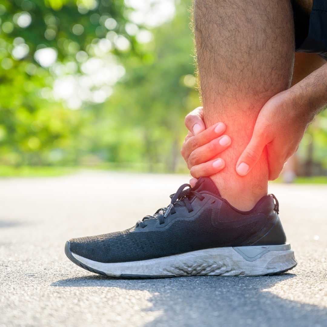
The runner who increases mileage too quickly. The gym-goer who suddenly adds daily sessions. The new military recruit who goes from sedentary to intensive training in two weeks. The badminton player who returns to daily court sessions after months off. These are the patients who develop stress fractures — and most of them spend weeks assuming it is just muscle soreness before they finally seek a proper evaluation.
Stress fractures are not dramatic injuries. There is no fall, no collision, no single moment of pain. They develop quietly, through the accumulation of repetitive mechanical load that exceeds what the bone can absorb and repair. By the time the pain becomes persistent enough to seek help, the fracture is often already several weeks old.
What Is a Stress Fracture?
Bone is living tissue that constantly remodels itself in response to mechanical load. When load increases gradually, bone adapts — becoming denser and stronger over time. When load increases faster than the bone can remodel — too much activity, too quickly, with too little recovery — microscopic cracks develop and accumulate faster than they are repaired. These cracks are stress fractures.
The distinction from a traumatic fracture is important. A traumatic fracture happens in a single moment from a significant force. A stress fracture develops over days to weeks from repetitive forces that would be entirely harmless individually — it is the cumulative effect, not any single impact, that breaks the bone.
Who Gets Stress Fractures?
Runners
Particularly those increasing weekly mileage too rapidly, switching surfaces suddenly, or running in worn footwear. Most common overuse injury in long-distance running.
Gym and Fitness Enthusiasts
Sudden return to intensive training after a break, rapidly increasing high-impact exercise volume (jumping, skipping, box jumps), or training through pain.
Military Recruits
Classic population — transition from low activity to intensive drill and marching over a short period. Tibial and metatarsal stress fractures are extremely common in early recruit training.
Court Sport Players
Badminton, basketball, volleyball, and squash players — repeated jumping and landing loads the foot and shin significantly.
People with Low Bone Density
Osteoporosis, low vitamin D, calcium deficiency, or eating disorders increase stress fracture risk at lower activity levels. Even normal walking can cause stress fractures in severely deficient bone.
Women with Menstrual Irregularity
The female athlete triad — low energy availability, menstrual irregularity, and low bone density — significantly increases stress fracture risk. Oestrogen plays a key role in bone health.
Symptoms — How a Stress Fracture Presents
Activity-Related Pain
Pain that begins during exercise and eases with rest — the defining early pattern. Initially present only at peak exertion, then progressively earlier in the session.
Point Tenderness
A specific, precise spot on the bone that is exquisitely painful to touch — one of the most reliable clinical signs of a stress fracture.
Progressive Worsening
Unlike muscle soreness that improves with days of rest, stress fracture pain worsens progressively over weeks if activity continues.
Mild Swelling
Localised swelling around the fracture site, often subtle and easily mistaken for a soft tissue problem.
Pain at Rest
In later stages, pain is present during normal walking or even at night — indicating the fracture is progressing.
No Clear Injury
The patient cannot recall any specific fall, twist, or trauma — just increasing pain with a specific activity over recent weeks.
Common Locations — Where Stress Fractures Occur
Metatarsals (Foot)
2nd and 3rd metatarsals most frequently. Classic in runners and military recruits. Usually manageable with rest and activity modification.
Tibia (Shin)
Most common stress fracture overall. Upper and lower thirds are higher risk. Pain along the inside of the shin bone during running. Requires careful management.
Fibula
Lower fibula most often affected. Generally lower risk of complications than tibia. Managed with activity modification and protected weight-bearing.
Navicular (Foot)
One of the most serious stress fractures — poor blood supply means healing is slow and unreliable. Often requires non-weight-bearing in a cast. Surgical fixation in some cases. Must not be missed.
Femoral Neck (Hip)
Serious stress fracture carrying risk of complete fracture and avascular necrosis if not treated promptly. Causes groin pain in active individuals. Requires urgent evaluation and often surgical fixation.
Calcaneus (Heel)
Heel bone stress fracture — painful with weight-bearing, specific tenderness when squeezing the heel from the sides. Common in military and endurance athletes.
Why Standard X-Ray Often Misses It
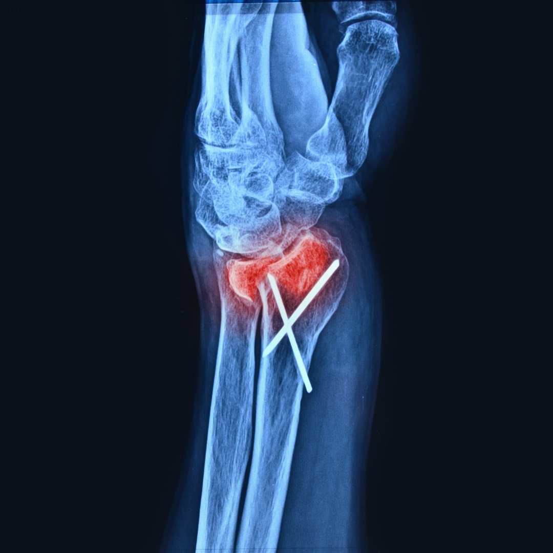
One of the most common frustrations for patients with stress fractures is being told their X-ray is normal — and being sent home without a clear answer. This happens because standard X-rays do not reliably detect stress fractures in the early weeks. The fracture line is too small to see, and the reactive bone changes that become visible later have not yet developed.
Diagnostic approach at Pure Ortho Hospitals, Sainikpuri
- Clinical history — activity type, training load change, timeline of pain development
- Physical examination — point tenderness over bone, hop test, fulcrum test for femoral shaft
- X-ray — performed but often normal in early stress fractures; may show periosteal reaction or fracture line after 2-3 weeks
- MRI — the investigation of choice; detects bone stress reaction and stress fractures from the earliest stage, determines severity, identifies high-risk fractures
- Bone scan — used when MRI is not available; sensitive but less specific than MRI
- Bone density assessment — particularly in younger women, patients with multiple stress fractures, or those with low-load fractures
Treatment — From Rest to Return to Sport
Treatment pathway at Pure Ortho Hospitals, Sainikpuri
- Activity modification — immediate reduction or cessation of the loading activity that caused the fracture; continuing to load a stress fracture risks complete fracture
- Protected weight-bearing — crutches or a walking boot for lower limb fractures, depending on site and severity
- Cross-training during recovery — swimming and cycling maintain fitness without bone loading while the fracture heals
- Bone health optimisation — calcium, vitamin D, and nutritional assessment; addressing any underlying deficiency that contributed
- Gradual return to activity — structured, progressive loading programme once symptoms have resolved and imaging confirms healing
- Surgical fixation — for high-risk fractures (femoral neck, navicular, certain tibial fractures) that require internal fixation to ensure healing and prevent complete fracture
Return to Activity — The Staged Approach
Rest Phase
Full activity cessation. Pain-free with normal walking. Typically 2-4 weeks.
Cross-Training
Swimming or cycling. No impact loading. Maintain fitness during healing.
Walking Programme
Progressive walking distances. Pain-free throughout. Usually week 4-6.
Return to Running
Walk-run intervals, increasing run time gradually. Typically week 6-10.
Full Activity
Return to full training and sport. MRI confirmation of healing before high-risk sport return.
Seek Urgent Evaluation For
- Groin or hip pain in a runner or active individual — possible femoral neck stress fracture
- Top of the foot pain without a clear injury that has not improved with 2 weeks of rest
- Any bone pain that is worsening despite stopping the aggravating activity
- Second or third stress fracture — warrants bone density and nutritional assessment
Femoral neck stress fractures in particular are time-sensitive. Call Pure Ortho Hospitals, Sainikpuri: 8686868208
Meet the Specialists at Pure Ortho Hospitals
Dr. G. Uday Sekhar Reddy
MBBS, MS Ortho, MCh Ortho
Dr. V.S. Abhilash Kumar S
MBBS, MS Ortho, FIJR, FISS (S.Korea, USA) — Clinical Director
Dr. Sai Karthikeya Badri
MBBS, D. Ortho, DNB
Dr. L. Sreeram
MPT (Ortho), FDOR, MIAP
Dr. L. Sri Dharani
BPT, MIAP, PTOTA (Canada)
Frequently Asked Questions
Other Departments at Pure Ortho Hospitals
Bone Pain That Builds During Activity Needs Proper Evaluation
Stress fractures caught early heal faster and rarely need surgery. Left untreated, they progress to complete fractures. Visit Pure Ortho Hospitals, Sainikpuri, Hyderabad for an MRI-based assessment.
Call 8686868208More from Pure Ortho Hospitals
This article is for patient education only. Please consult a qualified orthopaedic surgeon before making any treatment decisions.